
Skin cancer is the most common form of cancer
Overview
Like other forms of cancer, skin cancer results from the unrestrained growth and division of unhealthy cells. Perhaps because of our love for the sun, skin cancer is now the most common form of cancer and rates are increasing every year.
Skin cancer is categorized as one of two general types, melanoma and non-melanoma. Both types are principally caused by excessive exposure to the sun’s ultraviolet rays along with a genetic predisposition. People who have light skin, blue eyes and light hair, such as red or blond are more predisposed to developing skin cancer.
Prevention
The best way to treat a skin cancer is to prevent it from developing in the first place!
- Wear sunscreen with SPF 40 or higher, even in the winter
- Apply sunscreen 30 minutes before going outside, and reapply regularly throughout the day, especially when spending long periods in the sun. Don’t forget places like the back of your neck and your ears!
- Seek shade whenever you can
- Wear a wide-brimmed hat and UV-rated sunglasses that block the sun well
- Make sure your children do the same! Skin cancers start with sunburns in our childhood
One of the biggest risk factors for developing skin cancer later in life is suffering from bad sunburns in your youth. You can’t change what you did in your younger years, but you can help your children and loved ones avoid future skin cancers by instilling good sun habits in them now!
Catch Skin Cancer Early
Skin cancers are best treated early – when they are still small, thin, and are unlikely to have spread to other body sites.
Check your skin regularly, using a full body mirror or a partner to help see the places you can’t, to look for abnormalities such as:
- spots and sores that itch, crust or bleed
- open sores that do not heal within three weeks
- moles or beauty marks that change color or texture, have an irregular shape, are growing, or are new.
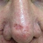
Basal Cell Cancer
Basal Cell Cancer (BCC) is the most common form of skin cancer, and fortunately is also the safest. It generally does not spread to lymph nodes or other parts of the body, and is generally not a threat to your life. However, it can be very destructive to the tissues of the body site where it is located if it is not treated properly.
This type of skin cancer fortunately is the least dangerous as it almost never spreads to other parts of the body. However, it must be treated since it will continue to grow, invading and destroying surrounding skin tissue, and can become very disfiguring.
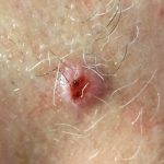
What does it look like?
Basal cell skin cancers usually appear on sun-exposed areas such as the face and neck, trunk, arms and legs.
The appearance of this type of skin cancer can vary. The early warning signs to look for are:
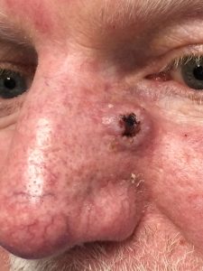
- A firm, flesh coloured or slightly reddish bump, often with a pearly border. It may have small blood vessels on the surface which gives it a red colour.
- A sore or pimple-like growth that bleeds, crusts over and then reappears. Any sore that does not heal within four weeks should be examined by your dermatologist.
- A small, red scaling patch.
What causes it?
Ultraviolet radiation from the sun is the prime cause of this skin cancer. Frequent severe sunburns and intense sun exposure in childhood increase the risk of developing BCCs in adulthood.
Studies have shown that fair-skinned people with blond or red hair and light skin that burns easily when out in the sun are at highest risk. BCCs usually start to appear as people get older, especially over age 50. However, these tumours are sometimes seen even in teenagers and in people in their early twenties. Organ transplant patients whose immune systems are compromised also face a higher risk.
How is it treated?
Treatment options depend on the tumour (size, location etc.) and the patient’s own health status. Treatment options may include surgical excision, shave and desiccation, cryotherapy, and/or radiation. Your doctor will help decide which treatment(s) are best for your cancer.
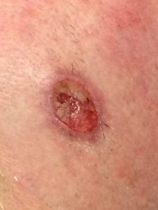
Squamous Cell Cancer
Squamous Cell Cancer (SCC) is the second most common form. SCC can also be very destructive to local tissues. However, it is more worrisome than BCC because it can sometimes spread to lymph nodes or other organs in the body if it is not treated. When this happens, it can sometimes be life-threatening.
This form of skin cancer must be treated because the lesion may continue to grow in size, damaging surrounding tissue, and may spread to other areas of the body.
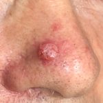
What does it look like?
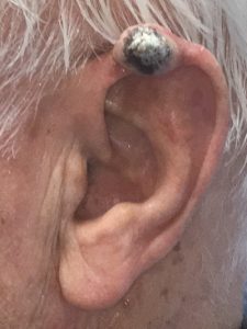
SCC appears on chronically sun-exposed areas such as the head and neck, arm, back of the hand and leg. SCC can be particularly aggressive when involving the ear or the lip.
Squamous cell skin cancers appear as thickened, red, scaly bumps or wart-like growths. They may also look like an open sore or crusted skin. This type of skin cancer may grow quickly over a period of a few weeks.

What causes it?
Frequent and cumulative sun exposure is the leading cause of this disease.
People who have suppressed immune systems or are taking anti-rejection drugs are also at higher risk.
How is it treated?
The treatment of this skin cancer consists mainly of surgical excision or radiation.
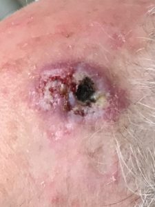
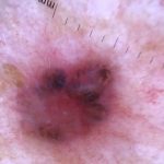
Melanoma
Melanoma is one of the rarest, but also one of the most dangerous forms of skin cancer. It has the highest rate of spread to other organs and carries the highest fatality rate of all the skin cancers. Outcomes can be dramatically improved by catching and treating melanoma early.
When found at an early stage, melanoma has a very high cure rate at more than 90 per cent. However, more advanced forms start to invade more deeply into the skin, where they can get into the blood stream or the lymphatic system. If this happens, it has a chance to spread to other parts of the body and may even cause death.
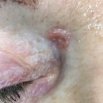
What does it look like?
Melanoma can develop in weeks or months, or take years. It can appear as a new mole or freckle-like spot on the skin, or in an existing mole that has started to change.
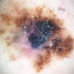
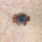
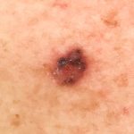
Know the ABCDE features suspicious for melanoma:
- Asymmetry – is the shape different from one side to the other?
- Border – are the edges irregular?
- Colour – are there multiple colours in the lesion?
- Diameter – is the spot getting bigger?
- Evolution – is the spot changing in any way: new, growing in size, changing shape or colour, becoming itchy or bleeding?
Also remember the Ugly Duckling Sign: Most moles on a person’s body look similar. A mole that looks different from all the others may be a melanoma. If you have a spot that looks or even feels different, or changes differently compared to other moles on the body, it should be checked by your dermatologist or family doctor as soon as possible.
Finally, while the features described here are the most common presentation for melanomas, not all melanomas look this way. In fact, sometimes the most dangerous ones have no brown colour at all – these are fortunately also the most rare. Therefore, any NEW or CHANGING skin lesions that don’t go away after a few weeks should be checked by your doctor.
What causes it?
Excessive exposure to ultraviolet (UV) from the sun and tanning beds plays a leading role in the development of melanoma and is the most preventable cause of this disease. Experts estimate about 90% of melanomas are associated with severe UV exposure and sunburns over a lifetime.
Other features that put people at higher risk of developing melanoma include:
- Light skin, fair hair and eyes, and lots of freckles
- Lots of moles on your body
- Personal history of having a melanoma already
- A close family member has had melanoma
The more risk factors you have, the higher your risk. However, even people with no risk factors and those with darker skin can also sometimes develop melanoma.
How do I protect myself?
The best ways to protect yourself are to:
- Know the risk factors above
- Learn the early signs of melanoma described above
- Check your skin monthly using a full length mirror, or get a partner to check those tricky-to-see places for you
- Be sun-smart all year round – see our tips under SKIN CARE
- Avoid tanning beds.
- Consult a doctor if you see any suspicious spots.
Remember, early detection and treatment is directly linked to a high survival rate.
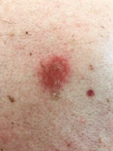
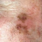
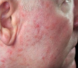
Actinic Keratoses
Although actinic keratoses (AKs) are not true skin cancers, it is important to have these lesions treated as they have the potential to change into squamous cell skin cancers.
What do they look like?
AKs appear on sun-exposed areas such as the face, ears, scalp, neck/chest, back of the hand, forearm and leg. People can have one, a few, or many at a time.
They can appear as:
- Red, rough, scaling spots, which may sting or itch
- Horny nodules, especially on the ears
- Dry, cracked or scaling skin along the edge of the lower lip that doesn’t heal
Skin Cancer Treatment
The most effective way to remove suspicious growths is with surgery. This can usually be done as a same-day procedure using local anesthesia, or in the operating room under general anesthesia.
In the clinic, growths may also be removed by cauterization, freezing, or cutting out. Other treatments may include radiation, lasers, or topical medications.
Your doctor will recommend the appropriate treatment(s) for you and help you through the decision making process. These include:
Surgery
Surgery to cut out the skin cancer is the most common and usually the most effective way of treating skin cancers, especially advanced ones.
Surgery is done by anesthetizing the skin in the area, and then using one of several surgical techniques to excise the cancerous tissue. These include:
- Local Excision: this type of surgery can be performed either with the patient awake, with local anesthesia, or with the patient asleep in the operating room, under general anesthesia. The surgeon cuts out all visible skin cancer tissue, along with a small cuff of “normal” appearing skin, to try to ensure any “fingers” of cancer cells that may extend from the main tumor are caught and removed as well. The wound is repaired and the excised skin is sent to the lab for analysis over 2-3 weeks to ensure all of the cancer was removed. This form of surgery has a cure rate of 90-95% for primary skin cancers, and 75-85% for recurrent cancers.
- Local Excision with Frozen Section Margins: this method is identical to Local Excision, except that a small rim of extra tissue is sent to the lab immediately for analysis at the time of excision. The lab usually processes and analyzes the tissue over 1-2 hours, then reports back whether or not any cancer remains to be cut out. This technique is most useful for recurrent cancers or ones that are hard to see with the naked eye, and can increase the cure rate of Local Excision to >95%. However, it requires a lot of expensive hospital resources and therefore is used only in special circumstances.
- Mohs surgery is a special form of skin cancer surgery performed by specially-trained dermatologists. The Mohs surgeon cuts out all visible skin cancer tissue, along with a small cuff of “normal” appearing skin, similar to Local Excision. The wound is then bandaged and the patient waits while the tissue is processed, and the Mohs surgeon him- or herself then analyzes the edges of the tissue to see if any cancer remains to be cut out. If cancer remains, the Mohs surgeon continues cutting out and analyzing more tissue specimens until the specimens are clear of any sign of cancer. This can sometimes take multiple rounds and several hours of surgery before the wound is clear of cancer and can then be repaired. Mohs surgery offers the highest cure rates of >95%. However, it also requires the most time, expense and hospital resources of all the surgical options, so wait times to see a Mohs surgeon in Canada can be very long.
Cryotherapy
Cryotherapy involves using liquid nitrogen in spray form to freeze unwanted skin tissue. It can be used for benign skin lesions like warts, skin tags, and seborrheic keratoses, or for some skin cancers.
After application of liquid nitrogen spray, the treated area of skin first becomes red and inflamed, then scabs over, then heals with new, healthy skin. Sometimes several treatments are needed to achieve complete removal of the lesion. The new, healed skin may be paler than the surrounding skin.
For more information about Cryotherapy, see the Cryotherapy Information Sheet.
Shave Excision/Biopsy
Shave excision is usually used to biopsy areas of skin that are suspicious for skin cancer. It is also used sometimes to treat select early and pre-cancerous skin lesions.
Shave excision involves using a blade to make a very shallow cut into the skin and “scrape” off the suspicious skin; usually the base of the wound is then cauterized to prevent bleeding and kill off any remaining pre-cancer cells.
The sample of suspicious skin is then sent to the lab for analysis. If the lab finds pre-cancerous tissue, usually no further treatment is required. However if advanced cancer is found in the sample, further surgery is usually required to make sure it is fully removed.
For more information, see Biopsy Information Sheet.
Prescription Creams
Several medicated creams are available for treatment of widespread sun damage and early skin cancers. These include Imiquimod (“Aldara”), 5-FU (“Effudex”), and others.
How they work: The creams activate your body’s natural immune defenses, helping your body to target and kill off sun-damaged skin cells in and around the areas where the cream is applied.
The creams must be applied for several weeks to be effective. Because they activate the immune system to attack the cancerous cells, there are several effects and side effects that you may experience during treatment:
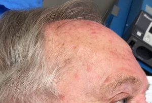
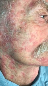
- You may experience no effects for the first few days of use as the cream ramps up your immune responses.
- Once ramped up, your immune system targets the sun-damaged skin cells. This can cause the skin to become very red, swollen, crusted, blistered and tender in and around where the cream is applied.
- After a few weeks, the sun-damaged cells are cleared, the the inflammation subsides, and the skin heals with fresh, smooth, healthy tissue.
Generally, the worse the inflammation experienced during use, the better the cream is working to kill off sun-damaged skin cells!
Prescription creams like Imiquimod can clear up to 80% of pre-cancerous and sun-damaged skin cells, and up to 50% of early skin cancers.
These creams should only be used under the guidance of your doctor. Be sure to follow application instructions carefully.
Radiation
Radiation is occasionally recommended for skin cancer treatment. It can be useful when:
- Surgery is not possible (due to very widespread disease, poor patient health, or tumor location)
- Surgery is refused
- Cancer remains even after surgery
- The cancer has very aggressive pathological features (invasion into nerves, bone, lymph tissue, blood vessels, etc.)
Radiation has a cure rate of about 80% for some types of skin cancer.
However, there are many potential reasons to avoid radiation:
- Radiation can only be given to an area ONCE in your lifetime. If you are at risk of getting another cancer in a similar area in your lifetime, radiation may not be a good choice now.
- Radiation may cause new cancers to appear 10-20 years later. If you are young, you risk developing new radiation-induced cancers later in your life.
- Radiation requires daily trips to the hospital over several weeks. This can be difficult for many patients to coordinate and tolerate.
- Radiation damages both diseased and healthy tissue in the area: this can result in unwanted side-effects depending on the location of the body that is treated (skin inflammation, dry mouth, hearing loss, visual damage, etc).